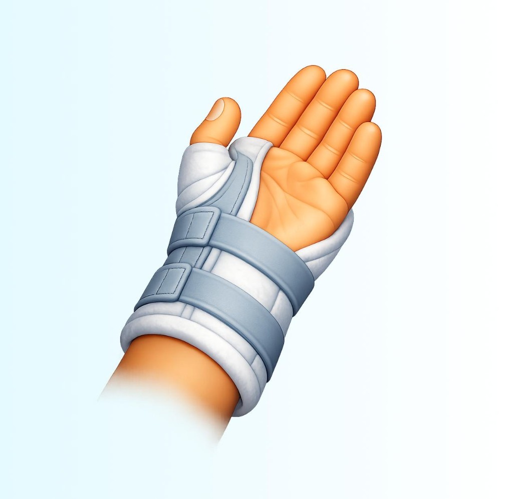
Wrist arthroscopy
A minimally invasive wrist procedure using a camera and small instruments inside the joint. It can be used to look for causes of wrist pain, smooth damaged tissue, or assist with certain repairs depending on the problem.
- Phases
3
- Red flagsEscalation
3
- Sources
1
Page reviewed — follow your clinical team for decisions.
Why it's done
- Persistent wrist symptoms when the diagnosis or treatment plan benefits from direct joint visualization
- Selected soft-tissue or cartilage problems treated arthroscopically when appropriate
Typical recovery phases
General patterns only — your protocol wins.
These phases describe common themes many teams use after this type of procedure. Your surgeon and hand therapist set the exact timeline, motion limits, and return-to-work or driving rules.
- Phase 1Days 0–10
Control swelling; protect portal sites.
Elevate the hand; move fingers often as allowed; keep dressings clean and dry.
- Phase 2Weeks 2–6
Restore comfortable wrist motion.
Gradually increase range and light use; avoid forceful twisting until cleared.
- Phase 3Weeks 6–12
Return toward usual activities.
Build grip and forearm endurance slowly; report new catching or locks.
Red flags — call your team
Contact your surgical team urgently for new or worsening symptoms like these. If you cannot reach them and the problem feels life-threatening, use local emergency services.
- Fever or rapidly spreading redness
- New numbness in several fingers
- Severe pain when elevating the hand
Sources
Independent references we used to shape this overview. They do not replace your clinician's instructions or your local emergency pathways.
- Wrist Arthroscopy — AAOS OrthoInfo(accessed 2026-04-30)